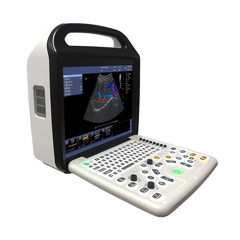
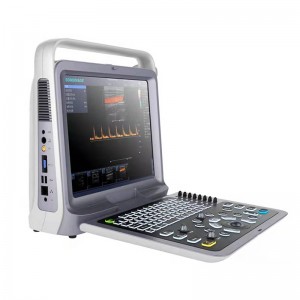
PET HOSPITAL USE P50 VETERINARY Color Ultrasonic Diagnostic Apparatus
★ Advanced imaging technology and superior image quality can provide fast and precise scans.
★ Applicable to scans of Equine, Bovine, Ovine, Swine, Feline, Canine, etc.
☆Applicable to different diagnosis of Abdomen, Obstetrics, Cardiology, small parts, vascular, tendon,etc
★ Comprehensive probe options can meet different clinic demands.
★ Powerful measurement software can provide comprehensive diagnostic bases.
★ Smart design and mobility make it easy to carry.
★ Built-in battery can support long-time outdoor diagnosis.
★ Efficient workflow can provide easy and comfortable operation experience.
A brand-new ultrasound diagnostic platform with Innovations in areas of digital electronics achieve a new level of ultrasound diagnostic precision and higher diagnostic confidence.
A revolutionary workflow control is provided with the user-centric architecture of the new software platform.
★Number of physical channels: ≥64
★Number of probe array element number: ≥128
Size:400mm(width) * 394mm(height) * 172mm(thickness)
Weight: machine weight is :
7.5kg(with no probe),
8.2kg (with one probe),
8.9 kg(with two probe)
15-inch, high resolution, progressive scan, Wide Angle of view
Resolution:1024*768 pixels
Image display area is 640*480
Internal 500GB hard drive for patient database management
Allow storage of patient studies that include images,clips,reports and measurements
Two active universal transducer ports that support standard(curved array, linear array), high-density transducers
156-pin connection
Unique industrial design provides easy access to all transducer ports
3C6C: 3.5MHz/R60/128, Convex array probe
7L4C: 7.5MHz/L38mm/128,Convex array probe
10L25C: 10MHz/25mm/128, Convex array probe
6C15C: 6.5MHz/R15/128,Micro convex array probe;;
3C20C: 3.5MHz/R20/128,Micro convex array probe;
★6I7C: 6MHz/L64mm/128,Intrarectal Linear array probe;
★5P2F: 5.0MHz/L10mm/64 Phased array probe;
Sector: selectable field of view (FOV) from 34 to 151degrees
Steerable Linear: variable steering angles for CFM and PW Doppler
Zoom:
- available on live, 2B, 4B and reviewed images
- up to 10X zoom
B-mode: Fundamental and Tissue harmonic imaging
Color Flow Mapping (Color)
Power Doppler Imaging (PDI)
PW Doppler
M-mode
B/M:Fundamental wave,≥3; harmonic wave: ≥2
Color/PDI: ≥2
PW: ≥2
B mode: ≥5000 frames
B+Color/B+PDI mode: ≥2300 frames
M, PW: ≥ 190s
Available on live, 2B, 4B and reviewed images
Up to 10X zoom
Format:
BMP, JPG, FRM(single image);
CIN, AVI(multiple images)
Support DICOM, conform to DICOM3.0 standard
★Built in workstation,support patient data search and browse

